
 Q. No.
Presence of intercalated discs help cardiac muscles to act as a functional syncytium. This is possible because the structure of these discs have:
1.
Tight junctions
2.
Gap junctions
3.
Adhering junctions
4.
Plasmodesmata


To unlock all the explanations of 38 chapters you need to be enrolled in MasterClass Course.
To unlock all the explanations of 38 chapters you need to be enrolled in MasterClass Course.

The type of epithelium that makes up the ‘Urothelium’ is:
1. Transitional
2. Stratified squamous non keratinized
3. Pseudostratified
4. Cuboidal brush bordered
Secretions from mammary glands are expelled by the contractile function of:
1. Smooth muscle
2. Skeletal muscle
3. Myoepithelium
4. Epithelio-muscular cell


In the given diagram of ultrastructure of a compact bone, identify A, B, C and D:
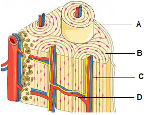
| 1. | Osteon, Lamellae, Haversian canal, Volkmann’s canal respectively |
| 2. | Lamellae, Osteon, Haversian canal, Volkmann’s canal respectively |
| 3. | Lamellae, Osteon, Volkmann’s canal, Haversian canal respectively |
| 4. | Osteon, Lamellae, Volkmann’s canal, Haversian canal respectively |
In the given diagram of areolar tissue, the structure that secretes mediators of inflammation is labeled by the letter:

| 1. A | 2. B |
| 3. C | 4. D |
The type of connective tissue shown in A and B in the given diagram are respectively located in:

1. Tendon and Ligament
2. Ligament and Tendon
3. Tendon and Skin
4. Skin and Tendon
To unlock all the explanations of 38 chapters you need to be enrolled in MasterClass Course.
To unlock all the explanations of 38 chapters you need to be enrolled in MasterClass Course.


Identify the incorrect statement regarding the parts labeled in the following diagram:

1. A is the perimysium
2. B is the structural unit of skeletal muscle
3. C is epimysium
4. D is the endomysium
The epithelium shown in the figure is present in the lining of:

1. Fallopian tubes
2. Trachea
3. Ureter
4. Thyroid follicles
The type of exocrine gland seen in the lining of gastric mucosa is shown by the figure:
| 1. | 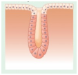 |
| 2. | 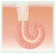 |
| 3. | 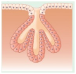 |
| 4. | 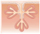 |


In the given diagram of a synovial joint, ligament and hyaline cartilage are represented respectively by the letters:

1. A and E
2. D and C
3. E and D
4. A and C
© 2026 GoodEd Technologies Pvt. Ltd.