
 Q. No.
The type of exocrine gland seen in the lining of gastric mucosa is shown by the figure:
1.
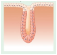
2.
3.
4.


In the given diagram of a synovial joint, ligament and hyaline cartilage are represented respectively by the letters:

1. A and E
2. D and C
3. E and D
4. A and C
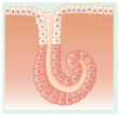
Based on the mode of secretion a gland shown in the following diagram will be called as:

1. Holocrine
2. Merocrine
3. Apocrine
4. Paracrine


The type of the neuron shown in the following diagram is seen in:

| 1. | Embryonic stages and Retina |
| 2. | Retina and Olfactory membrane |
| 3. | Retina and Dorsal root ganglion of spinal nerve |
| 4. | Olfactory membrane and Cerebellar peduncles |
In the given schematic diagram of ultrastructure of a myofibril, the functional unit of muscle contraction is shown by the letter:

| 1. | A | 2. | B |
| 3. | C | 4. | D |
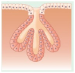
The following histology section depicts the structure of:

2. Single unit smooth muscle
3. Multi unit smooth muscle
4. Cardiac muscle

To unlock all the explanations of 37 chapters you need to be enrolled in MasterClass Course.
To unlock all the explanations of 37 chapters you need to be enrolled in MasterClass Course.


The calcium binding protein, troponin, is found in:
| 1. | Cardiac muscle and smooth muscle |
| 2. | Smooth muscle and skeletal muscle |
| 3. | Skeletal muscle and cardiac muscle |
| 4. | Skeletal muscle, smooth muscle and cardiac muscle |
What type of cell junctions are shown in the given diagram?

1. Tight junctions
2. Adhering junctions
3. Gap junctions
4. Desmosome
The disassembly of which of the following in uterine epithelial cells allows the blastocyst to penetrate between epithelial cells?
1. Tight junctions
2. Adhering junctions
3. Gap junctions
4. Apical cell membrane


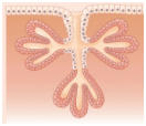
The cartilage shown in the given diagram is found in all the following locations except:
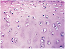
1. Articular cartilage
2. Larynx
3. Epiglottis
4. Ribs
© 2026 GoodEd Technologies Pvt. Ltd.